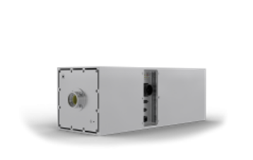
Detailed X-ray imaging requires clear visibility of small features, making focal spot size an important consideration. A smaller microfocus X-ray source can help reduce geometric unsharpness and support sharper image detail in precision inspection systems.
However, focal spot size works alongside the X-ray tube, high-voltage system, thermal management, beam geometry, and detector. Together, these elements influence the quality and effectiveness of the final image.
What Makes a Microfocus Source Different?
The defining characteristic of microfocus technology is its small focal spot. The focal spot is the area on the X-ray tube target where electrons strike to generate X-rays. When this area is reduced, the source can help limit the spread of the X-ray projection caused by the focal spot, which can improve the visibility of fine features.
For equipment manufacturers, this creates opportunities to design systems where image detail is an important requirement. The appropriate focal spot depends on the application, tube operating conditions, magnification, detector, and required resolution. A smaller focal spot should therefore be considered as part of the complete imaging architecture rather than as an isolated specification.
The Effect of Focal Spot Size on Image Detail
Focal spot size has a direct relationship with geometric sharpness. A smaller source area, such as that produced by a microfocus X-ray source, can reduce geometric blur, allowing edges and small structures to appear more clearly under suitable imaging conditions.
1. Reducing Geometric Unsharpness
Geometric unsharpness occurs when the finite size of the focal spot creates a blurred edge in the projected image. Reducing the focal spot can help limit this effect and improve image definition.
2. Revealing Fine Features
Detailed inspection often requires visibility of small structures or differences. A smaller focal spot can support the level of detail needed when the system is designed for precision imaging.
3. Supporting Component Inspection
Electronics and other small components can contain features that require careful examination. Microfocus sources can support equipment intended to inspect such details without relying solely on broad imaging conditions.
4. Improving Edge Definition
Sharp boundaries can make individual features easier to distinguish. A small focal spot can contribute to clearer edges when source-to-object and object-to-detector geometry are appropriately configured.
5. Working With Detector Geometry
Source performance does not work independently from the detector. Magnification, source-to-object distance, detector resolution, and focal spot size all interact to determine the final image characteristics.
Why Power and Focal Spot Need to Work Together
A smaller focal spot can support detailed imaging, but the source still needs an appropriate electrical operating range. High voltage and tube current determine how the X-ray tube operates and influence the amount of radiation produced. Therefore, source selection requires a balance between focal spot size and available output.
If the system requires higher output, the selected tube and power architecture need to accommodate those requirements without compromising the intended imaging conditions. The design should also consider heat generation, cooling, exposure time, and operating mode.
This balance is important for OEM systems because the best focal spot specification is not necessarily the smallest available. The appropriate choice depends on what the equipment needs to inspect or image and how the source will operate within the complete system.
Beam Configuration and System Design
Beam geometry also influences how an X-ray source can be incorporated into equipment. Different applications may use cone or fan beam configurations depending on the detector and inspection arrangement. Selecting the appropriate beam type can help align the source with the intended system layout.
Compact source construction can further support equipment development where internal space is limited. Integrated designs can combine the X-ray tube, high-voltage inverter, filament supply, and heat dissipation components within one unit. This can simplify the arrangement of important source functions within an OEM system.
Radiation shielding is another design consideration. Selected integrated source models include localized shielding, which can help address radiation management within the source assembly and reduce the need for additional shielding in the wider system.
Applications That Benefit From Detailed X-Ray Imaging
Microfocus technology can be valuable in applications where image clarity and small-feature visibility are important. Electronics inspection is one example, as X-ray systems can be used for component counting and examination of small elements. Industrial systems can also use high-resolution X-ray imaging for non-destructive testing and quality-related inspection.
The appropriate source depends on the equipment and application. Different products can provide different combinations of focal spot size, voltage, power, beam angle, cooling, shielding, and physical dimensions. This allows OEM developers to evaluate source technology according to the actual requirements of the system.
What OEM Developers Should Evaluate
Choosing a source for detailed imaging requires more than focusing on the smallest focal spot. The complete specification should be considered to ensure that the source can work effectively with the intended tube, detector, and equipment architecture.
- Focal spot size and required image resolution
- X-ray tube voltage and power requirements
- Tube current and intended operating range
- Cone or fan beam configuration
- Detector type and positioning
- Source-to-object and object-to-detector distances
- Cooling and thermal management requirements
- Physical size and mounting considerations
- Radiation shielding requirements
- Control and communication interfaces
These factors can help equipment developers determine whether a microfocus configuration is appropriate for a particular imaging objective. A balanced approach can prevent source selection from being based on one specification alone.
Building a Balanced High-Resolution System
A detailed X-ray image is the result of several coordinated elements. Source characteristics, focal spot size, electrical stability, beam geometry, detector capability, object positioning, and exposure conditions can all influence the final result. This means microfocus technology should be viewed as one part of a carefully engineered imaging system.
For OEM developers, an integrated source can provide advantages by bringing several essential functions together. Certain models combine high-voltage generation, tube operation, filament supply, and heat dissipation in a compact assembly. This approach can help simplify system integration while allowing manufacturers to select configurations suited to their equipment requirements.
See also: Expert Property Management Services in Northern Beaches
Conclusion
Microfocus technology matters for detailed X-ray imaging because a small focal spot can help reduce geometric unsharpness and improve the visibility of fine features. Its value becomes clearer when focal spot size is considered alongside voltage, power, beam geometry, detector performance, cooling, and overall system design.
For OEMs evaluating high-voltage power supply manufacturers, VJ X-Ray offers integrated X-ray source technology designed around different imaging and inspection requirements. Its range includes compact source configurations with small focal spots, high-voltage integration, thermal management, shielding, and application-focused designs, giving equipment developers options for building specialized X-ray systems.